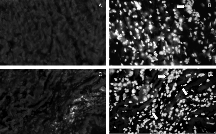
Figure 7.
Immunofluorescence detection of inflammatory IFN-γ-producing cells in paraffin-embedded heart sections taken from perforin (+/+) (a) and (−/−) (c) mice after 21 days of infection with Y strain T. cruzi. Nuclear labelling with DAPI corresponding to the same microscopic fields are also shown in (b) and (d), respectively. Arrows indicate inflammatory infiltrates. The original magnification was 630×.

