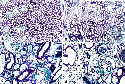
Figure 1.
Masson trichrome-stained histological sections from renal cortex (a, b, c and d) of a control rat (a) and of rats killed 5 (c) and 30 days (b and d) after glycerol injection. Note the presence of tubular dilatation, swelling and flattening of proximal tubular cells with brush border loss, diffuse interstitial oedema and interstitial inflammatory cellular infiltrates in (c) and of fibrosis, interstitial inflammatory cellular infiltrate and tubular dilatation and atrophy in (b) and (d) X120, (a) and (b); X280, (c) and (d).

