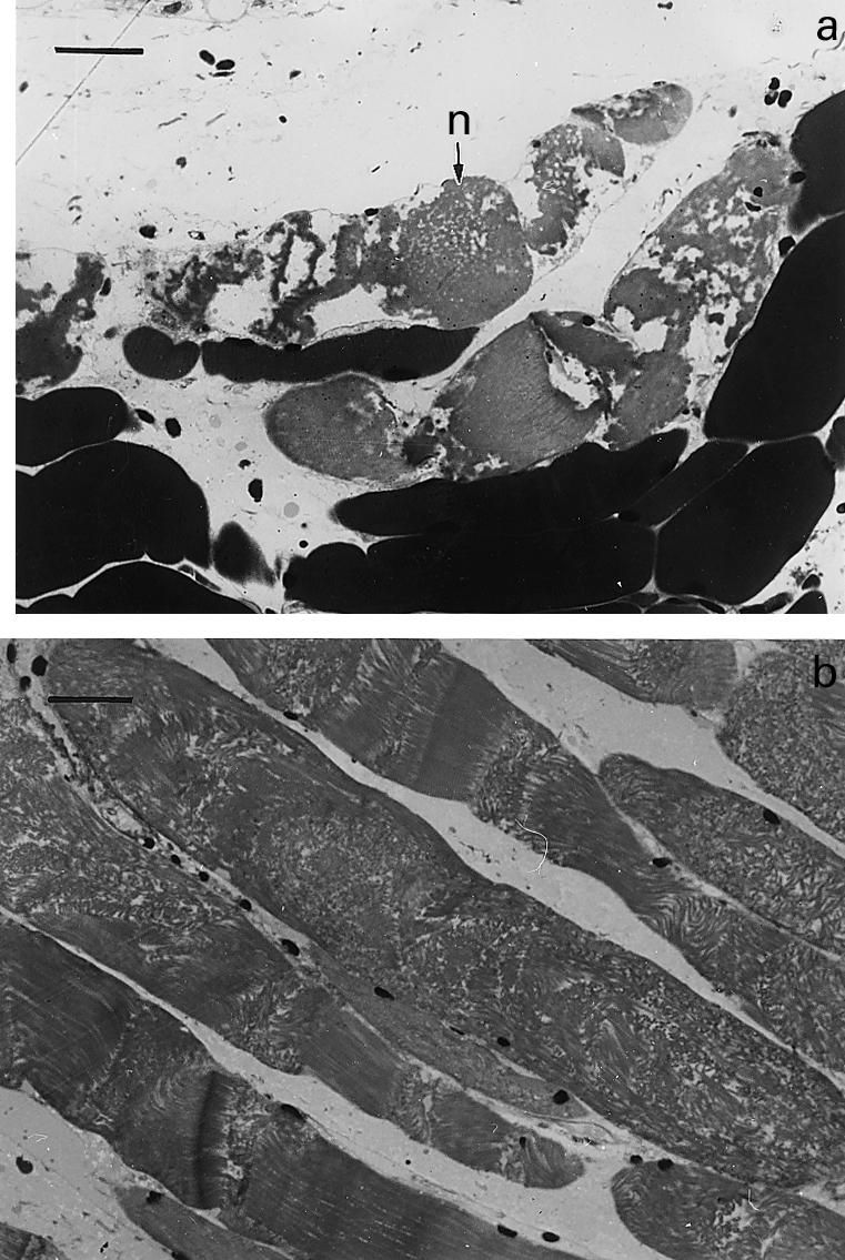
Figure 1.

Light micrographs of sections of gastrocnemius muscle taken 3 h after intramuscular injection of T. nattereri venom. (a) Necrotic muscle fibres (n) showing hypercontraction and clumping of myofilaments. Bar represents 25 µm (b) Group of necrotic fibres in which there is disorganization of myofibrillar structure with very few areas of hypercontraction. Bar represents 25 µm.
