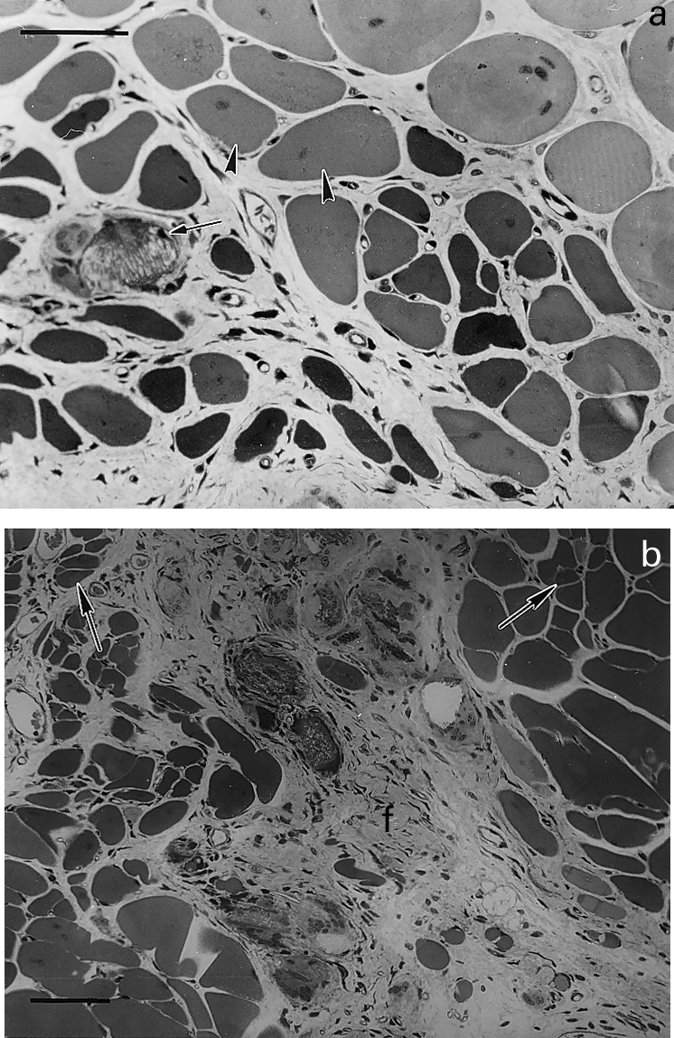
Figure 4.

Light micrographs of sections of gastrocnemius muscle taken at different times after intramuscular injection of T. nattereri venom. (a) 14 days; abundant regenerating fibres of varying size, some of them showing centrally located nuclei (arrow heads). Notice a calcified necrotic cell with a small regenerating fibre in its vicinity. Bar represents 50 µm (b) 28 days; portion of tissue in which regenerating fibres of varying size are observed, some of them showing fibre splitting (arrows). Prominent fibrosis (f) is observed in an area of poor muscle regeneration. Bar represents 50 µm.
