Figure 2.
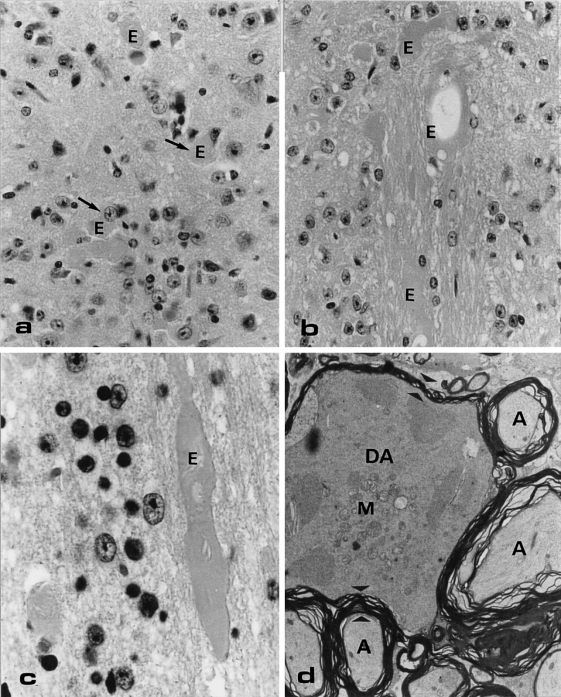
Light microscopical (LM) haematoxylin-eosine (HE)-stained section (a, b, c) and electron microscopical section (d) of abnormalities at the dorsal side of a transgenic mouse lumbar spinal cord. (a) Eosinophilic bodies/axonal swelling (E) scattered throughout the grey matter. Original Magnification (OM) = × 100. (b, c) Large eosinophilic bodies in the white matter, sometimes showing a torpedo-like shape (c) OM = 100x and × 250, respectively. (d) The degenerating axon (DA) in this electron micrograph is surrounded by normal axons (A). A clear difference in size and density of the cytoplasm and in number of mitochondria (M) in the degenerating and normal axon is shown. Note also the difference in thickness of the myelin surrounding the normal and degenerating axon (electron dense layered material in between the small arrowheads). OM = × 4.500.

