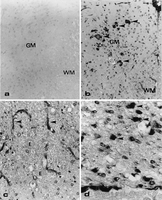
Figure 5.
Cross-section of the lumbar dorsal spinal cord of a wildtype (a) and transgenic mouse (b, c, and d), immunostained with anti-αA-crystallin. (a) No immunostaining is observed in the wildtype mouse. OM = × 50. (b) Various sized anti-αA-positive structures are present in the grey matter of the transgenic mouse. OM = × 50. (c) In the white matter of the transgenic mouse, small dots and stellate cells (arrows) are positive with anti-αA-crystallin. The eosinophilic bodies/axonal swellings (E) are not immunopositive. OM = × 100. GM, grey matter; WM, white matter. (d) The dorsal root shows many large dot-like immunopositive structures (arrowheads). OM = × 250.

