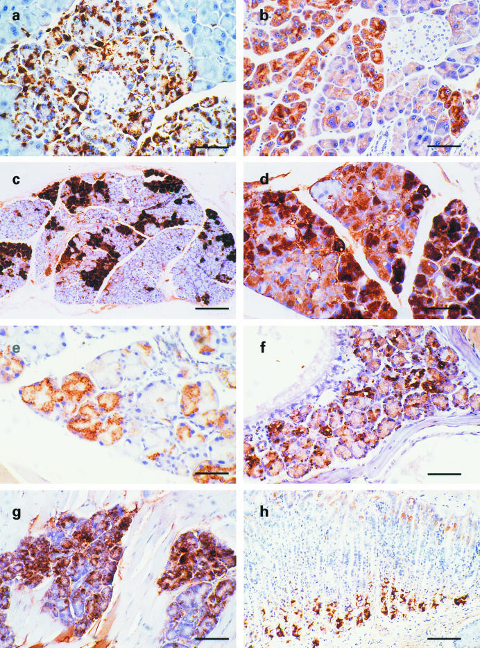
Figure 2.
Aβ-immunoreactive deposits in exocrine glands of 13592 mice. The pancreas and head of 13592 mice were stained with various antibodies using the avidin-biotin immunoperoxidase method. (a) Aβ-immunoreactive granular or globular deposits detected by the 4G8 antibody within acinar cells of a 7-month-old 13592 mouse; (b) A section adjacent to (a) was stained with the 994B antibody. The staining is found in the cytoplasm of acinar cells but is diffuse and less intense compared to (a); (c) Aβ-immunoreactive deposits visualized by the 6E10 antibody are patchy in this exorbital lacrimal gland from a 3-month-old 13592 mouse; (d) Aβ-immunoreactive granular or globular deposits detected by the 6E10 antibody in the intraorbital lacrimal gland of a 11-month-old 13592 mouse; (e) When stained with the 994B antibody, the cytoplasmic staining of a lacrimal gland from a 22-month-old 13592 mouse appears to be diffuse and lighter compared to that stained with antibodies against Aβ, such as 6E10 and 4G8; f-h are Aβ-immunoreactive deposits in (f) the DeSteno's gland of a 18-month-old 13592 mouse stained with the 6E10 antibody (g) the mucus glands in the tongue of a 11-month-old 13592 mouse stained with the 6E10 antibody, and (h) the gastric gland of a 24-month-old 13592 mouse stained with 4G8 antibody. Scale bars, 65 μm (a, b, and d), 205 μm (c), 50 μm (e and f), 75 μm (g) and 120 μm (h).

