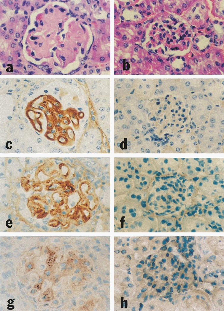
Figure 1.

Renal histology and immunohistochemistry of eight week-old IL-4 tg and wt mice. The kidney of an IL-4 tg animal (a) shows extensive glomerular hypertrophy and hypocellularity, basement membrane thickening and mesangial accumulation of eosinophilic material compared with the kidney of a wt littermate (b). The same tg mouse exhibits heavy glomerular Ig deposition (c); the control wt mouse kidney is negative for total mouse Ig staining (d). The expanded mesangium of the tg mouse kidney contains increased collagen type I (e), compared with the normal mouse kidney of the same age (f). TGF-β1 protein is increased in glomeruli of IL-4 tg kidneys (g); the kidney of a wt littermate displays low levels of glomerular TGF-β1 protein (h). a, b (H & E stain, ×290); c-h (immunoperoxidase staining, ×290).
