
Figure 3.
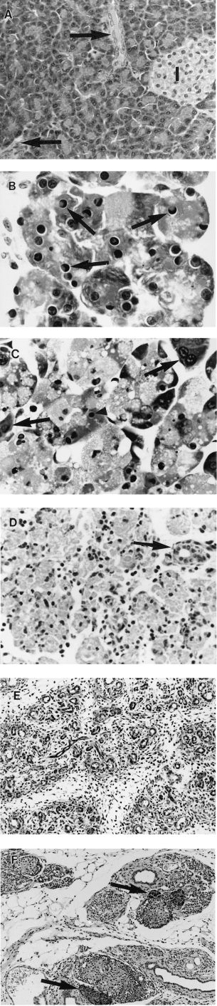
Pancreatic morphology after a single subcutaneous injection of saline or CHB. All H & E. (A) 48 h after saline. There is wide separation of ducts (arrows) and islets (I) by closely packed acinar cells (×360). (B) 12 h after CHB. Note numerous apoptotic acinar cells with characteristic nuclear morphology (arrows) (×1280). (C) 18 h after CHB. Most acinar cells contain pyknotic nuclear remnants (arrowheads) and show cytoplasmic swelling and vacuolation; a few appear normal (arrows) (×960). (D) 48 h after CHB. Advanced secondary necrosis affecting all acinar cells in field. Intact duct is indicated by arrow (×380). (E) 96 h after CHB. No acinar cells remain. Atrophic lobules comprise crowded ducts in a connective tissue stroma (×210). (F) 28 days after CHB. Sparse regenerative acini are seen adjacent to islets (arrows). Note few ducts in a collagenous stroma and prominent fatty infiltration (×210).