
Figure 4.
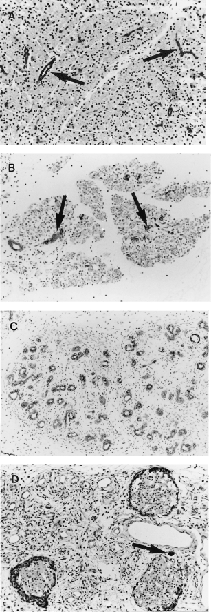
Pancreatic immunohistochemistry after a single subcutaneous injection of saline or CHB. All with haematoxylin counterstain. (A) 24 h after saline. Widely spaced keratin-positive ducts (arrows) are separated by closely packed keratin negative acinar cells (×170) (B) 48 h after CHB. Widely spaced keratin-positive ducts (arrows) separated by keratin-negative nonviable acinar cells (×190). (C) 96 h after CHB. Lobules comprise crowded keratin-positive ducts separated by loose connective tissue. No acinar cells are seen (×190). (D) 96 h after CHB. Ducts are negative for amylase. Note isolated amylase positive acinar cell (arrow) and periinsular amylase positivity (×170).