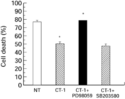
Figure 1.

CT-1 was cultured with cardiac myocytes for 30min under normoxic conditions in the absence ( ) and presence of 50μm PD98059 (▪) or 10μm SB203580 (
 ). No treatment (□). The inhibitors were added to the cells 30 min before the addition of CT-1. The CT-1 and inhibitors were removed from the cells which were then exposed to a 6-h simulated lethal hypoxic/ischaemic stress. The cells were harvested and cell death was assessed by trypan blue exclusion. *P < 0.05.
