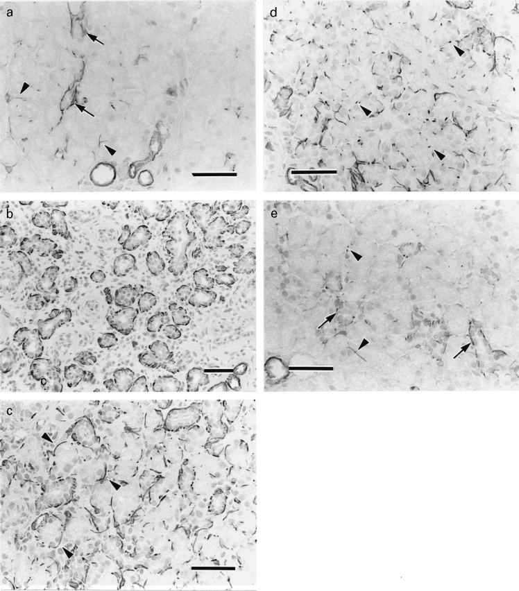
Figure 1.

Actin immunohistochemistry of a, the control and b–e, experimental glands. a, Actin-positive reaction is observed at the periphery of the intercalated ducts (arrows) and a few acini (arrowheads). Blood vessels are also positive. b, Day 0. Actin-positive reactions are identified around many residual ducts. c, Day 3. Newly formed acini are embraced by actin-positive cells (arrowheads). d, Day 10. Actin-positive reactions tend to be dots (arrowheads) around acini. e, Day 21. The positivity is observed at the periphery of intercalated ducts (arrows) and a few acini (arrowheads) (bar = 50 μm for all parts).
