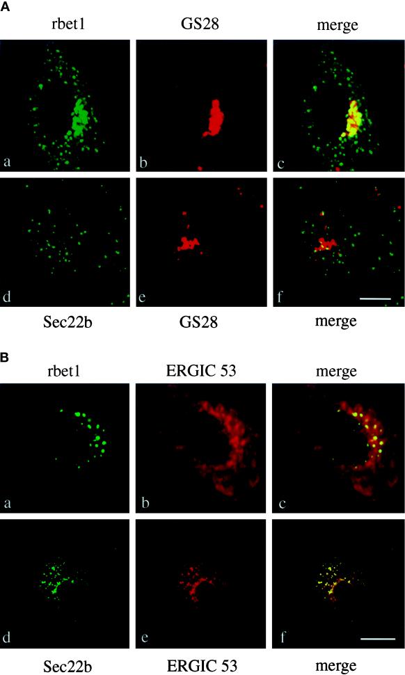
Figure 10.
(A) VSV ts045-infected Vero cells grown on coverslips were digitonin permeabilized and incubated at 32°C in complete transport cocktail buffer supplemented with either rbet1 or sec22b/ERS-24 antibodies for 120 min. Cells were fixed in paraformaldyhyde. Panels of inhibitory antibodies are shown in green. Panels of GS28 are shown in red. Also shown are the merged images. Bar, 5 μm. (B) VSV ts045-infected Vero cells grown on coverslips were digitonin permeabilized and incubated at 32°C in complete transport cocktail buffer supplemented with either rbet1 or sec22b/ERS-24 antibodies for 120 min. Cells were fixed in methanol. Panels of inhibitory antibodies are shown in green. Panels of ERGIC53 are shown in red. Also shown are the merged images. Bar, 5 μm.

