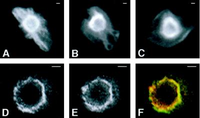
Figure 10.
Perinuclear AE1 anion exchangers and ankyrin are resistant to detergent extraction. Erythroid cells from a 10-d-old chicken embryo were extracted with 1% Triton X-100 in isotonic buffer before fixation in 1% paraformaldehyde in PBS. The cells were incubated with rabbit polyclonal antibodies specific for AE1 (A–C). Alternatively, cells were incubated with an AE1-specific mouse monoclonal antibody and a rabbit polyclonal antibody specific for ankyrin (D–F). Immunoreactive polypeptides were detected with DAR-IgG conjugated to lissamine and DAM-IgG conjugated to FITC and visualized on a Zeiss Axiophot microscope. The images showing the perinuclear localization of AE1 (D) and ankyrin (E) were pseudocolored green and red, respectively, and merged (F) in Adobe Photoshop. Erythroid cells preextracted with Triton X-100 before fixation exhibited reduced cell surface staining and strong perinuclear staining for both AE1 (A–D) and ankyrin (E). In addition, AE1 antibodies stained filamentous projections extending from the perinuclear compartment to the plasma membrane in some of the preextracted cells (A and B). Bar, 1 μm.

