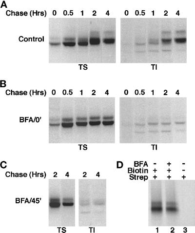
Figure 6.
Effect of BFA on the posttranslational processing of newly synthesized AE1 anion exchangers. Erythroid cells from a 10-d-old chicken embryo were pulsed for 15 min with 35S-Translabel and chased in complete media for various periods ranging from 30 min to 4 h (A). To identical cultures of cells, 10 μg BFA/ml were added either 20 min before the pulse (B), or after a 45-min chase (C), and the drug was included in the media for the duration of the experiment. At each time point, cells were detergent lysed and separated into soluble (TS) and insoluble (TI) fractions. Immunoprecipitates were prepared from both fractions using AE1-specific antibodies. Alternatively, 10-d erythroid cells were pulsed for 15 min with 35S-Translabel and chased in complete media for 1 h in the absence (D, lanes 1 and 3), or presence (D, lane 2) of 10 μg BFA/ml. At this time the cells were washed in Ringer’s buffer and incubated in the presence (D, lanes 1 and 2), or absence (D, lane 3) of 1 mg/ml NHS-SS-Biotin for 30 min at 4°C. The cells were then lysed in immunoprecipitation buffer, and immunoprecipitates were prepared using AE1-specific antibodies. After release from protein A agarose beads, biotinylated polypeptides were precipitated with streptavidin agarose. Each precipitate was analyzed on a 6% SDS polyacrylamide gel, and labeled anion exchangers were detected by fluorography.

