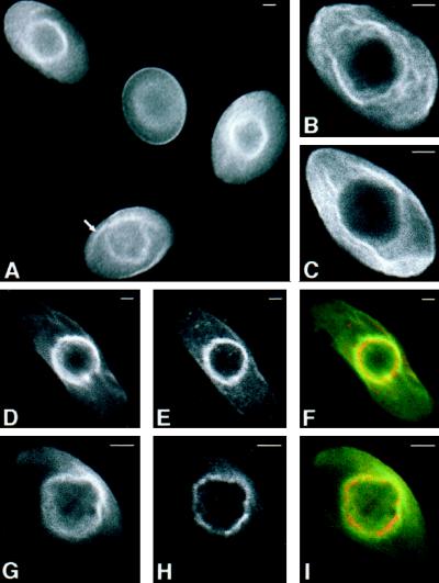
Figure 9.
Intracellular localization of chicken erythroid AE1 anion exchangers. Erythroid cells from a 10-d-old chicken embryo were fixed in 1% paraformaldehyde in PBS for 15 min. The cells were permeabilized with acetone and incubated with a rabbit polyclonal antibody specific for AE1 (A, B, C, and G). Alternatively, cells were incubated with an AE1-specific mouse monoclonal antibody and a rabbit polyclonal antibody specific for ankyrin (D–F). Immunoreactive polypeptides were detected with DAR-IgG conjugated to lissamine and DAM-IgG conjugated to FITC and visualized on a Zeiss Axiophot microscope (Carl Zeiss, Thornwood, NY) (A), or on a Bio-Rad laser scanning confocal microscope (B–I). After antibody incubations, the cell shown in panels G–I was incubated with 50 μg/ml NBD-ceramide for 1 h at 37oC, and washed. AE1 accumulated both in the plasma membrane and in a perinuclear compartment of erythroid cells (A). Filamentous membrane projections extend from this perinuclear compartment in some cells (marked by arrow in panel A). These filamentous membrane projections, shown at higher magnification in the 0.5-μm confocal images in panels B and C, do not appear to be present at the level of the cell surface. The 0.5-μm confocal images showing the perinuclear distribution of AE1 (D) and ankyrin (E) were pseudocolored green and red, respectively, and merged (F) in Adobe Photoshop. Likewise, the 0.5-μm confocal images showing the perinuclear distribution of AE1 (G) and NBD-ceramide (H) were pseudocolored green and red, respectively, and merged (I). Bar, 2 μm.

