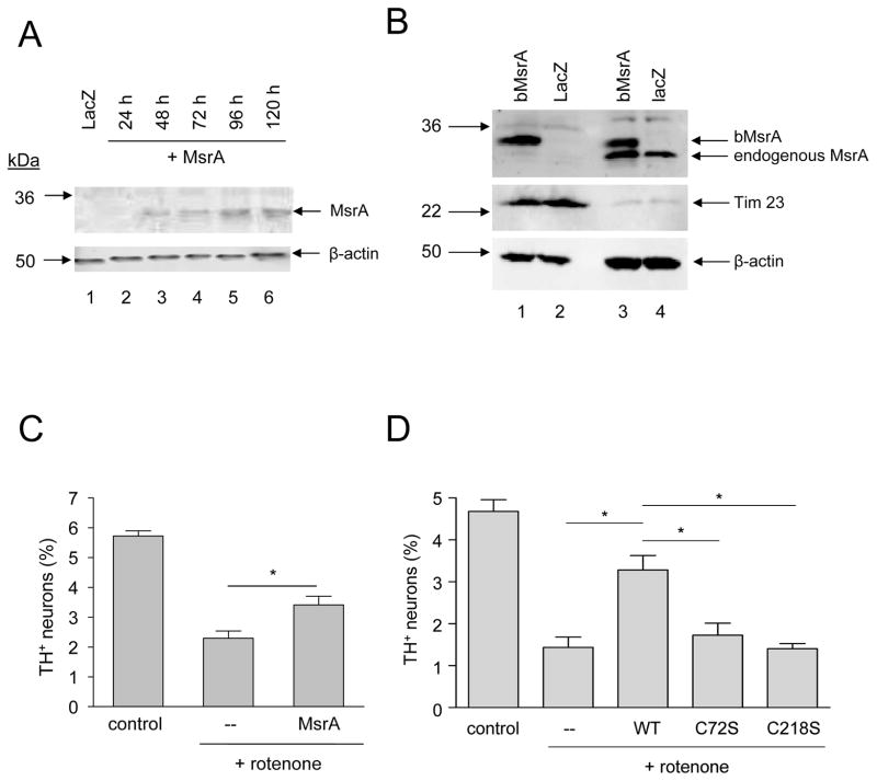
Figure 1.
MsrA protects primary dopaminergic neurons against rotenone toxicity. (A) Western blot analysis of MsrA expression levels in primary midbrain cultures. Lane 1: lysate from cells transduced with LacZ lentivirus for 72 h and then incubated in fresh media for 48 h; lanes 2–6: lysates from cells transduced with MsrA lentivirus for 24 h (lane 2), 48 h (lane 3), 72 h (lane 4), 72 h followed by a 24-h incubation in fresh media (lane 5), or 72 h followed by a 48-h incubation in fresh media (lane 6). 12 μg of protein was loaded in each lane. (B) Western blot showing the distribution of MsrA between mitochondrial fractions (lanes 1 and 2) and cytosolic fractions (lanes 3 and 4). MES23.5 cells were transduced with adenovirus encoding bovine MsrA (bMsrA) (lanes 1 and 3) or LacZ (lanes 2 and 4). (C) Wild-type MsrA expressed from a lentiviral construct inhibits dopaminergic cell death induced by rotenone (48 h). (D) Wild-type MsrA (but not the catalytically inactive mutants C72S and C218S) expressed from an adenoviral construct suppresses neurotoxicity elicited by rotenone (24 h). The data in (C) and (D) are presented as the mean +/− SEM, N = 3; *p<0.05.

