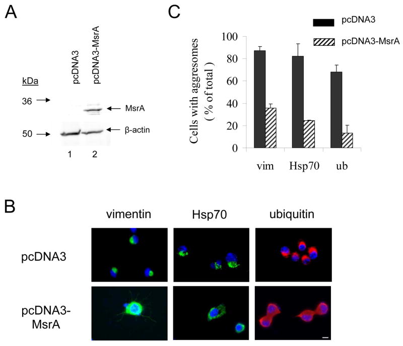
Figure 3.
MsrA suppresses aggresome formation induced by rotenone in MES23.5 cells. (A) Western blot analysis of MsrA expression levels in MES23.5 cells stably transfected with pcDNA3 (lane 1) or pcDNA3-MsrA (lane 2). (B) Immunocytochemical analysis of aggresome formation. MES23.5 cells stably transfected with pcDNA3 (top row) or pcDNA3-MsrA (bottom row) were treated with rotenone. The cells were stained with a primary antibody specific for vimentin (left column), Hsp70 (middle column), or ubiquitin (right column). Nuclei were stained with DAPI (blue). The scale bar corresponds to 10 μm. (C) Quantification of rotenone-induced aggresome formation in cells transfected with pcDNA3 or pcDNA3-MsrA and stained for vimentin (‘vim’), Hsp70, or ubiquitin (‘ub’). The data are expressed as the percentage of total cells with aggresomes (mean +/− SD, N = 3, p<0.001, Student’s t test for each stain).

