
Figure 8.
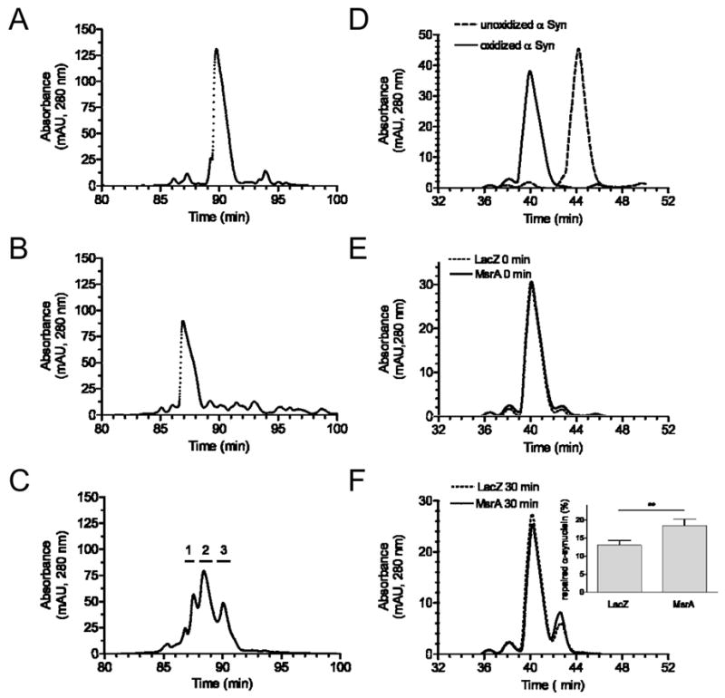
RP-HPLC chromatograms showing MsrA-mediated repair of recombinant α-synuclein. (A) Unoxidized α-synuclein. (B) α-Synuclein oxidized by incubation with H2O2. (C) Oxidized α-synuclein after incubation with recombinant MsrA (2 h, 37 °C). Horizontal lines above the chromatogram correspond to the collection times of fractions 1, 2, and 3. (D) Mixture of unoxidized and oxidized α-synuclein. (E) Oxidized α-synuclein after mixing with lysate of PC12 cells transduced with MsrA or LacZ adenovirus, followed by immediate termination of the reaction (i.e. ‘0 min’ incubation). (F) Oxidized α-synuclein after a 30-min incubation with lysate of PC12 cells transduced with MsrA or LacZ adenovirus. Inset: Quantification of the species eluting at ~42.5 min (corresponding to 3-MetSO α-synuclein) via peak area measurements. The data are expressed as the percentage of total α-synuclein converted to the 3-MetSO species (mean +/− SEM, N = 3, *p<0.05, Student’s t test). Note that different elution gradients were used in (A)-(C) versus (D)-(F).