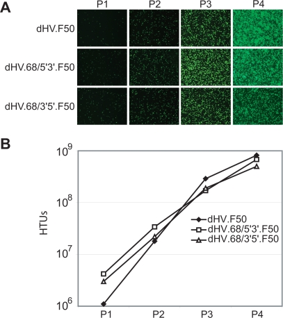
Figure 3. Propagation of rep68-positive and fiber-modified dHVs.
(A) Direct fluorescence microscopy analysis of the PER.tTA.Cre76 cells used for the serial propagation of dHV.F50, dHV.68/5′3′.F50 and dHV.68/3′5′.F50. (B) Flow cytometric analysis of HeLa cells employed for the determination of the eGFP transfer activity of clarified producer cell lysates derived from consecutive rounds of vector amplification. P1, passage 1; P2, passage 2; P3, passage 3; P4, passage 4.

