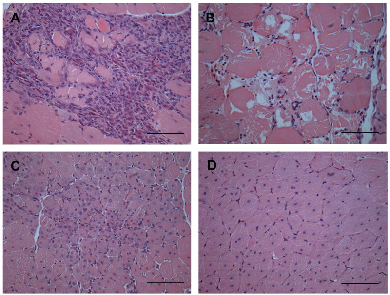
Fig 1. Histological features on a transverse section of exercised adult mdx quadriceps muscle stained with Haematoxylin and Eosin (all images are from the same muscle section).
A) Active muscle necrosis characterised by many inflammatory cells which have infiltrated dystrophic myofibres (sarcoplasm is barely visible). B) Active muscle necrosis characterized by fragmented sarcoplasm of dystrophic myofibres with irregular shape and few myonuclei; inflammatory cells are not conspicuous. C) Recent regeneration shown by small dystrophic myofibres (sometimes seen as smaller myotubes) with central nuclei. D) Regenerated muscle indicated by large mature dystrophic myofibres with central nuclei. Scale bar represents 100μm.

