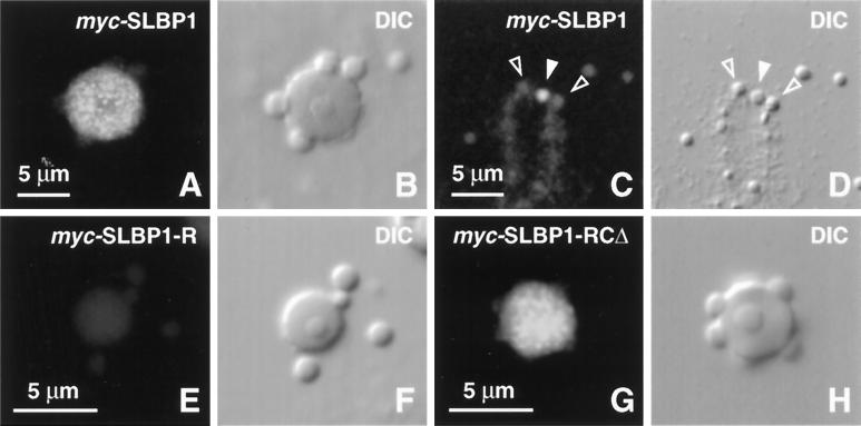
Figure 6.
Localization of myc plus NLS–tagged SLBP1 in GV spreads. Each pair of panels shows a DIC image and the corresponding immunofluorescent image after staining with mAb 9E10 against the c-myc tag. (A and B) A single coiled body from an oocyte injected 8 h previously with transcripts of full-length myc-SLBP1. The tagged protein accumulates in the matrix of the coiled body in a distinctly granular pattern. (C and D) End of a chromosome 8 h after a similar injection. The solid arrowhead points to a stained terminal granule; the open arrowheads point to two adjacent unstained B-snurposomes. (E and F) A single coiled body and three B-snurposomes 8 h after injection of transcripts of myc-SLBP1-R, which consists of the RNA-binding domain alone. Staining is at background level. (G and H) A single coiled body 8 h after injection of transcripts of myc-SLBP1-RCΔ. This protein accumulates in the matrix of the coiled body despite lacking a functional RNA-binding domain.

