Figure 9.
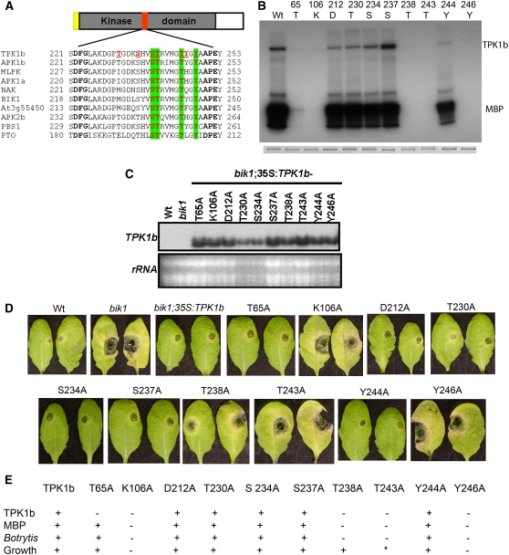
TPK1b Is a Functional Protein Kinase That Localizes to the Plasma Membrane.
(A) Alignment of the activation segment of TPK1b and related kinases.
(B) Autophosphorylation and MBP phosphorylation activities of GST-TPK1b and its mutant derivatives in vitro. Top panel, autoradiogram of the gel; bottom panel, Coomassie blue staining.
(C) RNA blot showing transgenic expression of TPK1b substitution mutants in the Arabidopsis bik1 mutant.
(D) Responses of bik1 plants expressing TPK1b and its substitution mutants to A. brassicicola inoculation.
(E) Summary of kinase activity, Botrytis response, and growth-related phenotypes of the Arabidopsis bik1 expressing TPK1b wild type and substitution mutants.
In (A), TPK1b activation segment phosphorylatable residues are in red and underlined. Residues that are phosphorylatable and conserved in TPK1b-related kinases are shaded green. In (D), A. brassicicola disease symptoms are from 4 DAI. In (E), the asterisk indicates partial complementation; +, rescues phenotypes or shows auto (TPK1b) or MBP phosphorylation activity; −, fails to rescue the bik1 growth phenotype or shows no TPK1b or MBP phosphorylation activity.

