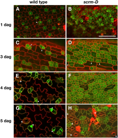
Figure 2.
Time Sequence of Stomatal Differentiation in scrm-D.
Images of the abaxial epidermis of wild-type and scrm-D cotyledons. TMM:GUS-GFP (green) was used to monitor stomatal lineage cells. Arrowheads, GFP positive cells underwent cell division; asterisks, meristemoids; +, mature stomata. Images were taken under the same magnification. Bar = 50 μm.
(A) Wild-type cotyledon at 1 d after germination (dag).
(B) scrm-D cotyledon at 1 d after germination.
(C) Wild-type cotyledon at 3 d after germination.
(D) scrm-D cotyledon at 3 d after germination.
(E) Wild-type cotyledon at 4 d after germination.
(F) scrm-D cotyledon at 4 d after germination.
(G) Wild-type cotyledon at 5 d after germination.
(H) scrm-D cotyledon at 5 d after germination.

