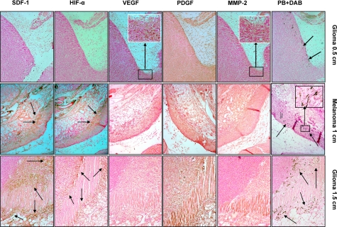
Figure 5.
Expression of different angiogenic and chemoattractant factors at the sites of migrated labeled AC133+ cells in tumors. Top row: expression of different angiogenic and chemoattractant factors at the sites of migrated labeled AC133+ cells in rat glioma at a tumor size of 0.5 cm (consecutive sections). Prussian blue staining shows migration of cells at the periphery of the tumor (arrows). MMP-2 and VEGF staining show expression of these factors at the corresponding sites of cell migration (inset, brown cells). PDGF staining shows generalized expression both in the tumor and adjacent surrounding tissues. HIF-1α and SDF-1 staining do not show expression of the factors at the corresponding sites of migrated cells. Middle row: expression of different angiogenic and chemoattractant factors at the sites of migrated labeled AC133+ cells in human melanoma at a tumor size of 1 cm (consecutive sections). Prussian blue staining shows migration of cells at the periphery of the tumor and at the sites of invasion into surrounding muscles and tissues (arrows). MMP-2 staining shows mild expression of MMP-2 throughout the tumor and surrounding tissues. PDGF staining also shows generalized expression both in the tumor and adjacent surrounding tissues. VEGF staining shows no localized expression of these factors at the corresponding sites of cell migration. HIF-1α and SDF-1 staining show very strong localized expression of the factors at the corresponding sites of migrated cells (arrows). Inset: multiple DAB enhanced Prussian blue positive cells. Bottom row: Expression of different angiogenic and chemoattractant factors at the sites of migrated labeled AC133+ cells in rat glioma at a tumor size of 1.5 cm (consecutive sections). Prussian blue staining shows migration of cells at the periphery of the tumor and at the sites of invasion into surrounding muscles and tissues (arrows). MMP-2 staining shows mild expression of MMP-2 throughout the tumor and surrounding tissues. PDGF staining shows generalized expression both in the tumor and adjacent surrounding tissues. VEGF staining shows no localized expression of these factors at the corresponding sites of cell migration. HIF-1α and SDF-1 staining show very strong localized expression of the factors at the corresponding sites of migrated cells (arrows). All images ×10.

