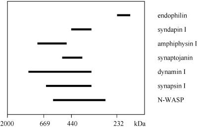
Figure 6.
Syndapin I exists in a high molecular weight complex. Rat brain high-speed supernatant was analyzed by gel filtration over Superose 6. Aliquots of each fraction were resolved on 6–15% SDS-PAGE and blotted to nitrocellulose. Proteins were detected by antibody reaction and overlay analyses with GST-Ddyn(PRD) and GST-SdpI, and the staining intensities of the fractions were plotted. Some proteins eluted in sharp peaks, whereas others were more broadly distributed. The horizontal bars end at the position at which the concentration of the protein in a fraction was half-maximal and give an estimate of the distribution. Half-maximum widths are shown for endophilin, syndapin I, amphiphysin I, synaptojanin, dynamin I, synapsin I, and N-WASP. Standards for column calibration correspond to dextran 2000 (2000 kDa), thyroglobulin (669 kDa), ferritin (440 kDa), and catalase (232 kDa).

