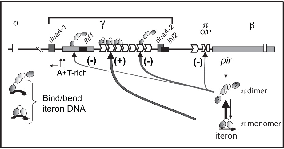
Figure 1.

Plasmid-encoded elements involved in the regulation of replication of γ ori plasmids. A physical map of the replicon of R6K showing the relative locations of the seven tandem iterons of γ ori, the single iteron and the half-iteron of α ori and β ori, respectively, the inverted half-repeats in the operator site adjacent to the pir gene, two DnaA sites, the integration host factor (IHF) site, the A+T-rich region of γ ori, and different paths of ori activation and inhibition by π monomers and dimers, respectively. Symbols representing monomers and dimers are labeled and the WH1 and WH2 recognition helices of both monomers and dimers are represented by ‘1’ and ‘2’, respectively. Shading indicates that two monomer subunits of a dimer make head-to-head contacts while two monomers bound to two tandem iterons are proposed to make head-to-tail contacts. A monomer contacts the iteron with both WH1 and WH224 while a dimer most likely contacts the iteron only with WH2 of one of the two subunits.
