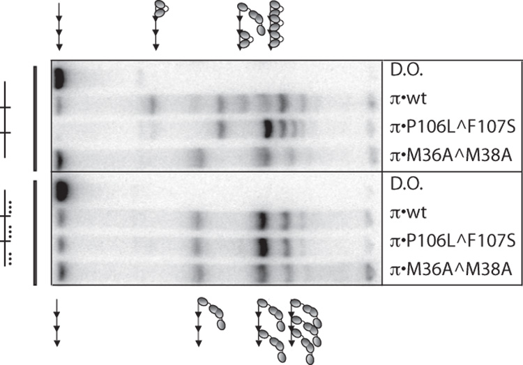
Figure 2.

In vitro binding patterns of π•wt to three tandem iterons with and without mutations in the “monomer-only” side of each iteron. Binding assays were performed with a probe containing three wt iterons in the left four lanes and three monomer-deficient iterons (mutations in the 14th, 17th, and 18th, bp) in the right four lanes. These iteron mutations are depicted by (…). π variants are labeled. DNA probe preparation and gel shift titrations were carried out exactly as previously described33 except that: 110 pg labeled iteron-containing probe was used in the binding reactions and Promega (Madison, WI) 6X loading dye was added prior to loading the gel. Probe sequences and construction are described in Table 1 of the Supplementary Materials. 200 ng of π was added to each binding reaction. His π•WT, His-π•P106L^F107S, and His-π•M36A^M38A were purified as described.38,39 D.O. is DNA only. Symbols representing monomers and dimers of π are the same as in Figure 1.
