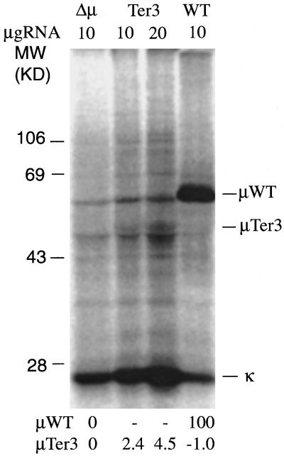
Figure 6.
In vitro translation of μ RNA. As described in MATERIALS AND METHODS, total RNA (10 or 20 μg) from the indicated cell lines was translated in vitro using rabbit reticulocyte lysate and [35S]methionine. The IgM-related material was then immunoprecipitated with rabbit anti-IgM serum and analyzed by SDS-PAGE. The intensity of the indicated μWT and μTer3 bands was quantified by PhosphorImager. The background for the Δμ cell line was subtracted, and the resulting values were then normalized to the value obtained for the μWT band. These normalized values are listed below each lane.

