Fig. 8.
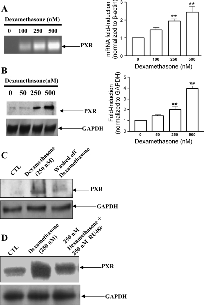
Dexamethasone stimulated increased transcript levels and protein expression of PXR in rat brain endothelial cells. Primary endothelial cells were treated with increasing concentrations of dexamethasone for 24 h. A: transcript levels of PXR were evaluated by RT-PCR, using total cellular RNA as described in materials and methods. **P < 0.01 vs. untreated cells. B: protein levels of PXR in nuclear extract were evaluated by immunoblotting using 25 μg protein loaded per lane. Representative autoradiograph from an experiment performed at least 3 times is shown. **P < 0.01 vs. untreated cells. C: representative Western blot demonstrating increased PXR expression in the presence of dexamethasone and reduced expression after 24 h without dexamethasone. D: representative Western blot demonstrating that glucocorticoid receptor antagonist RU486 partially inhibited the dexamethasone-induced stimulation of PXR.

