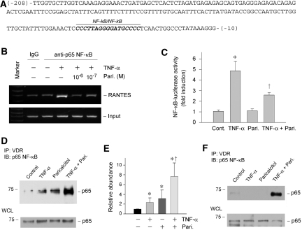
Figure 9.
Paricalcitol induces a physical association between VDR and p65 NF-κB, inhibits its binding to RANTES promoter, and represses NF-κB–mediated gene transcription. (A) Partial sequence of human RANTES gene promoter region. Highlighted is the NF-κB element in this region. (B) ChIP assay demonstrated that paricalcitol abrogated the binding of p65 to its cognate cis-acting element in the RANTES promoter. HKC-8 cells were treated with TNF-α and paricalcitol as indicated. (C) Luciferase reporter assay demonstrated that paricalcitol repressed the NF-κB–mediated gene transcription in response to TNF-α treatment. Relative luciferase activity (with control group = 1.0) is presented. Data are means ± SEM from three experiments. *P < 0.01 versus control; †P < 0.01 versus TNF-α treatment group. (D and E) Co-immunoprecipitation revealed that paricalcitol induced an increased binding of VDR to p65 NF-κB. HKC-8 cells were transfected with p65 NF-κB expression vector, followed by treatment without or with TNF-α, paricalcitol, or both as indicated. Cell lysates were then immunoprecipitated with anti-VDR antibody, followed by immunoblotting with anti-p65. Whole-cell lysates were also immunoblotted with anti-p65 for normalizing p65 abundance. Data from three independent experiments are shown (E). *P < 0.05 versus control; †P < 0.05 versus TNF-α or paricalcitol treated alone. (F) Paricalcitol also induced a physical interaction between VDR and endogenous p65 NF-κB. HKC-8 cells were treated without or with TNF-α, paricalcitol, or both and then assessed for endogenous p65/VDR complex formation by co-immunoprecipitation.

