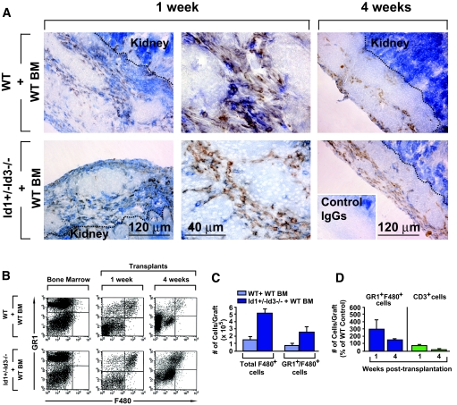
FIG. 3.
Detection of inflammatory leukocytes in the islet grafts. A: Tissue sections from islet grafts of bone marrow–reconstituted wild-type and Id1+/−Id3−/− mice, at 1 and 4 weeks after transplantation, stained by two-color immunohistochemistry for the pan-leukocyte marker CD45 (blue) and the myeloid marker F480 (brown) or control IgGs (inset). A leukocytic inflammatory infiltrate, comprising myeloid cells is apparent in the grafts from both experimental groups. The dotted lines mark the border of the grafts with the kidney. The intense blue staining in the kidney is background due to color development by the alkaline phosphatase endogenous to the kidney epithelium. Images are representative of n = 3 grafts per experimental group. B: Flow cytometric analysis of leukocytes isolated from the grafts at 1 and 4 weeks after transplantation stained by two-color immunofluorescence for the myeloid markers GR1 and F480. An increased percentage of GR1highF480+ cells in the graft of bone marrow–reconstituted Id1+/−Id3−/− mice is evident compared with wild-type controls. Theses cells are not present in the bone marrow of either mouse. The dot plots are representative of n = 4 experiments. C and D: Quantitative analysis of GR1highF480+ and CD3+ cell subsets detected by flow cytometry in the graft bone marrow–reconstituted Id1+/−Id3−/− and wild-type mice at 1 and 4 weeks after transplantation. Bars are means ± SE of n = 4 independent determinations. (Please see http://dx.doi.org/10.2337/db08-0244 for a high-quality digital representation of this image.)

