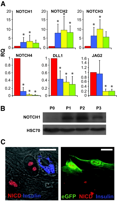
FIG. 2.
Upregulation of the NOTCH pathway in cultured human islet cells and eGFP+ cells derived from β-cells. A: qPCR analysis of RNA extracted from islet cells derived from eight donors at the indicated passage numbers. P indicates passage number and weeks in culture. RQ indicates relative quantification compared with P0. Data are means ± SD (n = 8). Asterisks indicate statistical significance, compared with P0 (P < 0.03 for NOTCH1; P < 0.04 for NOTCH2; P < 9 × 10⁻⁴ for NOTCH3; P < 0.03 for NOTCH4; P < 0.005 for DLL1; P < 0.026 for JAG2). The increase in NOTCH3 mRNA levels at P3 was marginally significant (P = 0.06). B: Immunoblotting for NOTCH1 120-kd transmembrane fragment in protein extracted from islet cells at the indicated passage number. HSC70 served as a loading control. C: Immunofluorescence analysis of islet cells (left panel) and eGFP+ cells derived from β-cells (right panel) following 10 days in culture. Bar = 20 μm. (Please see http://dx.doi.org/10.2337/db07-1323 for a high-quality digital representation of this figure.)

