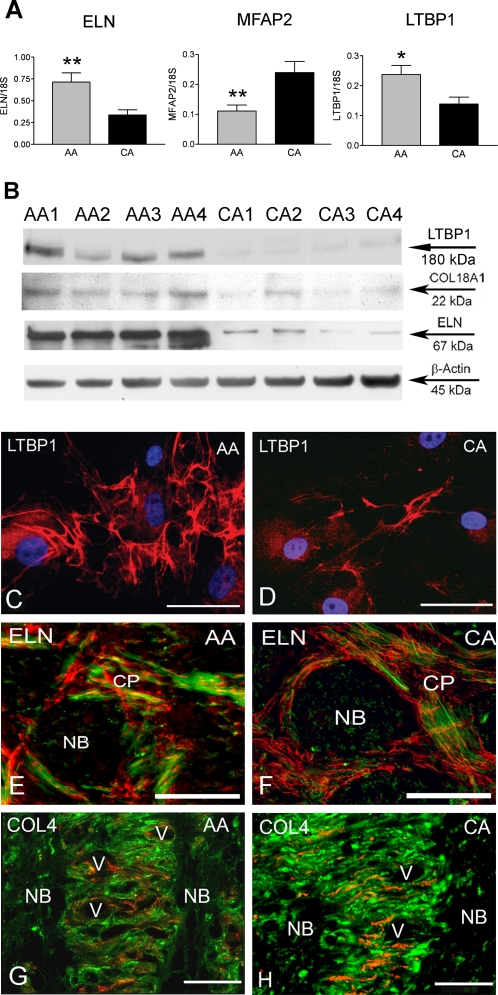
Figure 5. Differential expression of genes associated with the extracellular matrix (ECM) in AA astrocytes compared to CA astrocytes.
A. Confirmation of three differentially expressed adhesion genes by qRT-PCR in human normal ONH astrocytes: Elastin (ELN), microfibrillar-associated protein 2 (MFAP2) and latent transforming growth factor beta binding protein 1 (LTBP1). Genes were normalized to 18S. Graphical representation of the relative mRNA levels in normal AA and CA astrocytes (n = 8, respectively, two-tailed t-test was used. ** indicates p<0.01 and * indicate p<0.05). B. Representative Western blots of astrocyte cell lysates with LTBP1, ELN and collagen type XVIII antibodies. β-actin was used as a loading control. Note that AA1-4 donors express more LTBP1, ELN and collagen type XVIII than CA1-4 donors. C, D. Immunocytochemistry of LTBP1 in AA and CA astrocytes. LTBP1 (red) is more abundant in the cytoplasm and extracellular space in AA astrocytes compared with CA astrocytes. Nuclei stained with DAPI (blue). Magnification Bar: 25 µm. E, F. Double immunostaining of ELN (red) and GFAP (green) in representative cross sectional of the ONH tissue in AA and CA donors. ELN is located in the cribriform plates and not in the nerve bundles. Astrocytes cell bodies are located in the cribriform plates (CP) and extend processes into the nerve bundle (NB). Note that there are no apparent differences in ELN staining between AA and CA samples. E is from a 75 year-old AA male donor and F is from a 74 year-old CA female donor. Colocalization between ELN and GFAP in the AA tissue is microscopic effect showing the overlap between ELN and astrocytes processes. Magnification bar: 25 µm. G, H. Double immunostaining of collagen type IV (red) and GFAP (green) in representative sagittal sections of ONH tissues from AA and CA donors. Collagen type IV and GFAP follow the lamellar structure of the astrocytic basement membranes in the human lamina cribrosa. Note that staining for collagen type IV is more intense and abundant in the CA donor than in the AA donor. G is from a 65 year-old AA male donor and H is from a 57 year-old CA male donor. V: blood vessel, Magnification bar: 35 µm.

