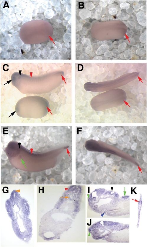
Figure 2. xPer1 is expressed from neural plate to late tailbud stages.
Shown are in situ hybridization results depicting expression of xPer1 mRNA. Panels A, B, D, and F show dorsal views of the embryos and panels C and E show side views of the embryos. All whole mount embryos (as well as panel I) are oriented with the anterior facing left. Dorsal is toward the top in all images. A and B show neural plate staining at stage 15/16 and stage 18, respectively. Red arrows denote posterior mesoderm staining (A–F). C and D show a neural tube stage embryo on the bottom (stage 22) and an early tailbud stage embryo on top (stage 33). The black arrow denotes eye expression and the red arrowhead shows somite staining. C and D also show xPer1 expression in the CNS and posterior mesoderm (red arrow), as well as the otic vesicle (C, black arrowhead). Panels E and F depict xPer1 expression in the CNS, somites (red arrowhead), otic vesicle (black arrowhead), pronephric tubules (green arrow) and posterior mesoderm (red arrow) in a late tailbud stage embryo. Panels G–J show sections of late tailbud embryos (G–H and J are transverse sections and I is a sagittal section). G shows expression in the neural tube, retina, lens, and pineal gland (orange arrowhead). H shows expression in the notochord (orange arrow) and the somites (red arrowhead). In panel I, the olfactory pit (green arrowhead), otic vesicle (black arrowhead), heart (blue arrowhead) and pronephros (green arrow) were stained. Panel J shows olfactory pit staining (green arrowhead). Panel K shows posterior mesoderm staining in the tail tip (red arrowhead).

