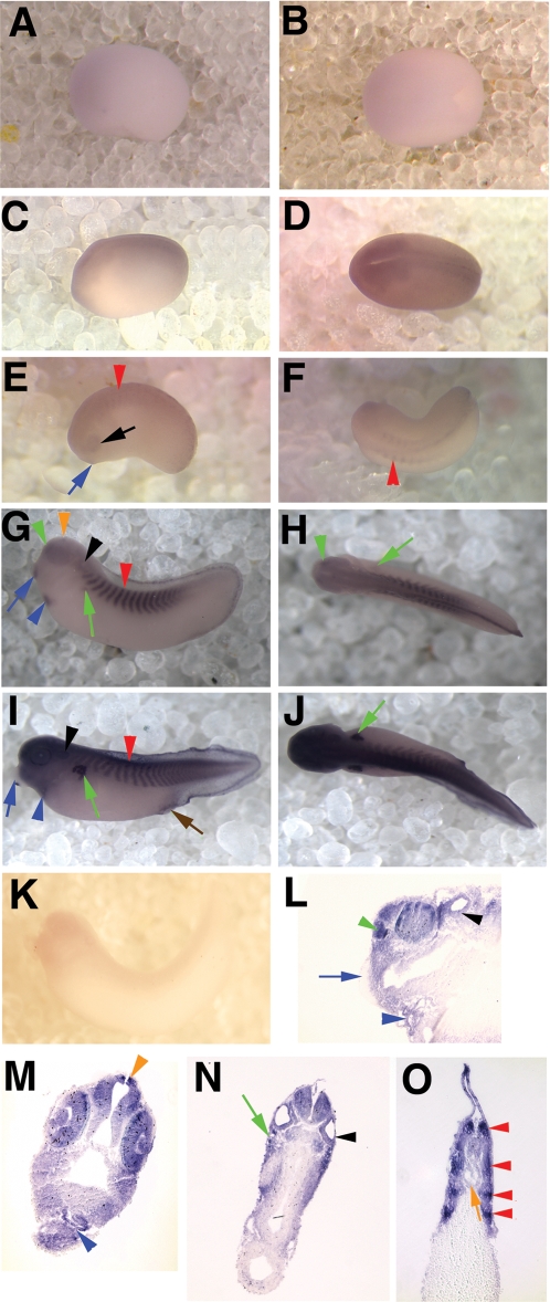
Figure 5. xNocturnin is expressed from neural plate to late tailbud stages.
Shown are in situ hybridization results depicting expression of xNocturnin mRNA. All embryos in this figure are shown with the anterior facing left. Side views of the embryos are depicted in panels A,C,E,G,I, and K and dorsal views in panels B,D,F, H, and J. Low levels of xNocturnin were first detected in the neural plate of stage 15/16 embryos A and B. C and D show neural plate staining in a stage 18 embryo. E and F show a neural tube stage embryo (stage 24) with xNocturnin expression in the eyes (black arrow), somites (red arrowhead), and cement gland (blue arrow). G and H show early tailbud stage embryos with staining in the otic vesicle (black arrowhead), pronephric tubules (green arrow), heart (blue arrowhead), olfactory pit (green arrowhead), pineal (orange arrowhead), cement gland (blue arrow) and somites (red arrowhead). Late tailbud stages (I and J; stage 39) show similar results but additional staining in the anus/blastopore (brown arrow) and cement gland staining is absent (blue arrow). Sagittal (L) and transverse sections (M–O) of late tailbud embryos confirm xNocturnin expression in the brain, retina and lens (M), otic vesicle (N, black arrowhead), olfactory pit (L, green arrow), pronephric tubules (N, green arrow), heart (M, blue arrowhead), notochord (O, orange arrow) and in the somites (O, red arrowheads). xNocturnin is absent from the cement gland at late tailbud stages (L, blue arrow). No expression was seen using a sense probe specific to Nocturnin (K).

