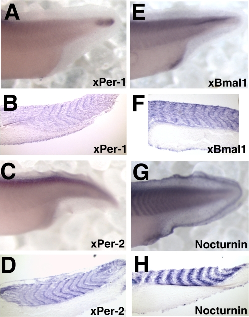
Figure 6. A comparison of somite staining in the posterior of late tailbud embryos (stage 36–38).
Shown are in situ hybridization results depicting RNA expression in paired whole mount and sagittal sections of the posterior of embryos stained with xPer1 (A–B), xPer2 (C–D), xBmal1 (E–F), and Nocturnin (G–H).

