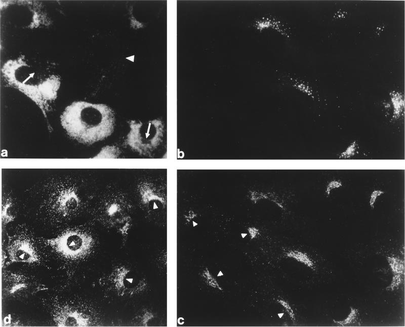
Figure 1.
Steady-state distribution of the AP-1 adaptor complex and clathrin in normal and MPR-negative cells in vivo. Normal and MPR-negative mouse embryo fibroblasts were cocultured on glass coverslips, fixed with formaldehyde, and then probed either with mAb 1D4B directed against mouse LAMP-1 (panel a), a mixture of affinity-purified rabbit antibodies against the CI-MPR (panel b), and a mAb (clone 41) recognizing the γ subunit of AP-1 (panel c). The MPR-negative cells are easily distinguished from the MPR-positive cells either by the presence of numerous swollen LAMP-1–positive structures (panel a) or by the lack of CI-MPR labeling (panel b). A control, MPR-containing cell is indicated (panel a, arrowhead) as is the region in some of the MPR-negative cells that is devoid of LAMP-1–positive structures and contains the bulk of the other organelles in these cells (panel a, arrows). Similar levels of AP-1 staining are observed in both the MPR-lacking (panel c, arrowheads) and the normal cells. A culture of the MPR-negative cells alone was processed for analysis with mAb X22, directed against the clathrin heavy chain (panel d). The concentration of clathrin over the juxtanuclear region of each cell is clearly visible.

