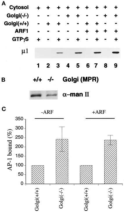
Figure 4.
Recruitment of AP-1 onto MPR-positive and MPR-negative Golgi-enriched membranes. (A) Recruitment assays containing 50 μg/ml normal (+/+) or MPR-negative (−/−) Golgi-enriched membranes, 5 mg/ml gel-filtered rat liver cytosol, 4 μM recombinant myristoylated ARF1, and 100 μM GTPγS were prepared on ice as indicated. After incubation at 37°C for 15 min, the Golgi-enriched membrane pellets were recovered, resolved on polyacrylamide gels, and transferred to nitrocellulose. The blot was probed with the anti–μ1-subunit antibody, RY/1. Only the relevant portion of the blot is shown. (B) Aliquots of 10 μg protein of the Golgi-enriched membrane fractions derived from either normal (+/+) or the MPR-negative (−/−) cells used in panel A were analyzed by immunoblotting with the polyclonal anti–α-mannosidase II antiserum. (C) The μ1-subunit signal from six (−ARF) or four (+ARF) independent experiments was quantitated by densitometry and the average values (± SEM) are expressed relative to the Golgi marker content of the membranes. The values for the MPR-positive membranes were arbitrarily set to 100%.

