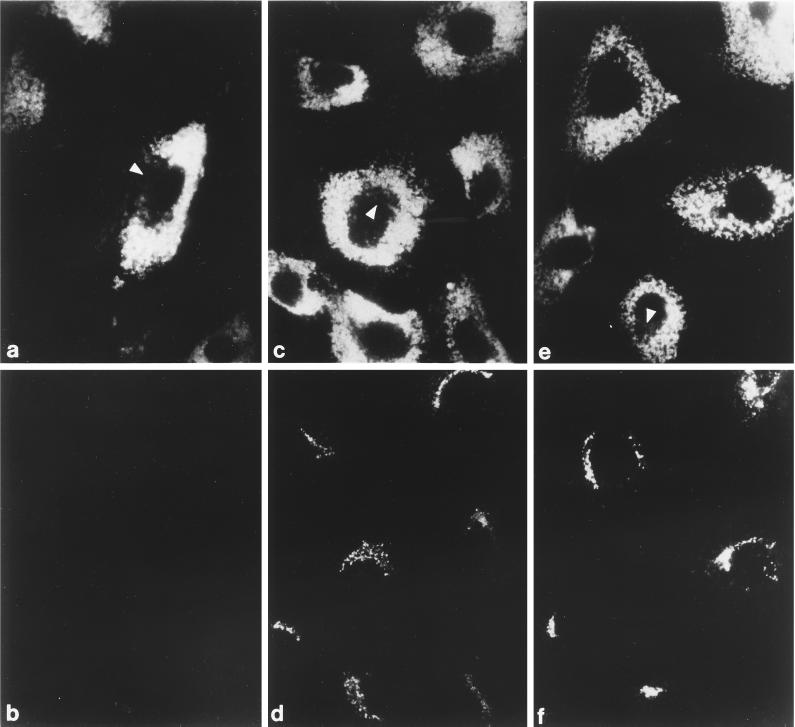
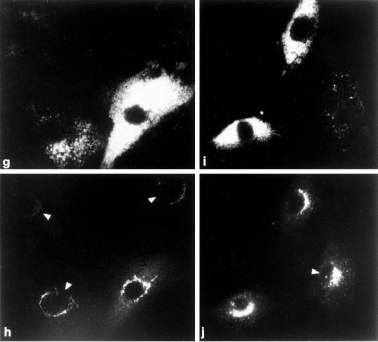
Figure 5.
Recruitment of AP-1 in permeabilized MPR-negative fibroblasts. (a–f) Digitonin-permeabilized receptor-negative cells were incubated at 37°C for 20 min with ∼5 mg/ml gel-filtered rat liver cytosol and either 1 mM GDP (panels a and b) or 100 μM GTPγS (panels c–f). After washing, the cells were fixed and then prepared for indirect immunofluorescence using a mixture of the anti–LAMP-1 mAb 1D4B (panels a and c) and affinity purified anti–γ-subunit antibody AE/1 (panels b and d) or mAb 1D4B (panel e) and affinity purified anti–β-subunit antibody GD/1 (panel f). The conditions for photography and printing of panels b, d, and f were identical. In some cells, the region that is devoid of LAMP-1–positive structures containing the bulk of the other perinuclear organelles is indicated (panels a, c, and e, arrowheads). (g–j) Cocultures of MPR-positive and MPR-negative fibroblasts were permeabilized, mixed with 5 mg/ml gel-filtered cytosol and 100 μM GTPγS, and incubated at 37°C for 20 min. After washing the cells were fixed and prepared for immunofluorescence analysis using a mixture of the anti–LAMP-1 mAb 1D4B (panels g and i) and affinity-purified anti–γ-subunit antibody AE/1 (panels h and j). Two representative images are shown, and the normal, MPR-positive cells are indicated by the arrowheads.


