
Figure 2.
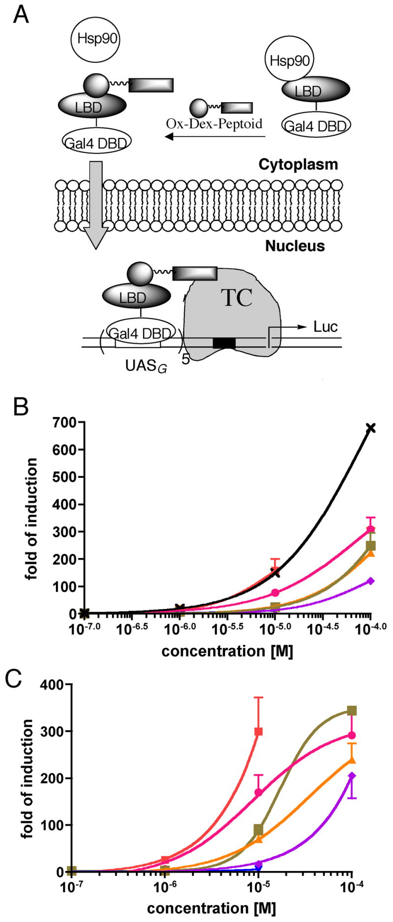
Cellular activities of OxDex-BHK1-6 (see Figure S3 for structures). (A) Cartoon of the assay employed to measure the AD-like activity of the peptoid (see text for details). TC = transcription complex. (B) HeLa cell permeability of the OxDex-peptoid conjugates. OxDex-AEEA-CONH2 (×), OxDex-BHK1 (
 ), OxDex-BHK2(
 ), OxDex-BHK3(
 ), OxDex-BHK4(
 ), OxDex-BHK5(
 ) or OxDex-BHK6(
 ). This assay31 is almost identical to that shown in A, except that the Gal4 fusion protein also contains a VP16 AD. The values shown are ratios of the level of induction measured using dual-luciferase assay system (Promega; Gal4-responsive/constitutive). Data points for OxDex-BHK1 and OxDex-BHK3 at 100 μM could not be obtained due to significant cytotoxicity at this concentration. (C) Activation domain-like activity of OxDex-BHK1-6 in the assay shown schematically in (A). The symbol designations are the same as in (B). The data, shown as the mean + SD or mean - SD, are representative of at least three independent experiments with each point measured in duplicate. The curves are the best fit of the data to the sigmoidal dose-response model in Prism4.0.