Figure 4.
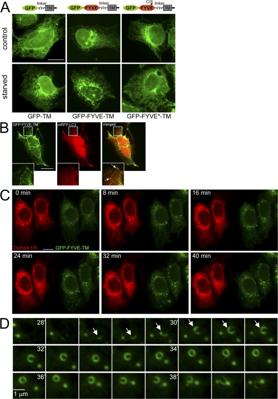
ER-anchored FYVE domain translocates to starvation-induced punctate structures in a pathway dependent on PI(3)P. (A) Three GFP-based constructs were fitted with a linker region, a transmembrane domain (TM), and a short cytoplasmic tail: GFP alone (GFP-TM), GFP fused to the WT FYVE domain of FENS-1 (GFP-TM-FYVE), and GFP fused to a point mutant of the FENS-1 FYVE domain unable to bind PI(3)P (GFP-FYVE*-TM). All three constructs were expressed transiently in HEK-293 cells, and their localization was examined with or without amino acid starvation for 60 min as indicated. Note that GFP-TM-FYVE translocated to punctate structures during starvation. (B) HEK-293 cells were transfected with GFP-FYVE-TM and mRFP-LC3 and starved for 60 min. Note that puncta of the two reporters colocalize, with the GFP construct frequently encircling mRFP-LC3 membranes (arrows). In general, cells had many more GFP-FYVE-TM than mRFP-LC3 puncta, and on average, 80% of LC3 puncta colocalized with a GFP-FYVE-TM particle. Insets show enlarged views of the boxed regions. (C) Live imaging of HEK-293 cells coexpressing GFP-FYVE-TM and dsRED-ER and starved for 60 min. Also see Video 1 (available at http://www.jcb.org/cgi/content/full/jcb.200803137/DC1). (D) A region indicated in C (bottom middle, box) is expanded and shown for 28–40 min during starvation. Note the formation and collapse of a ringlike particle. Arrows mark the particle at early stages. Bars: (A–C) 20 μm; (D) 1 μm.

