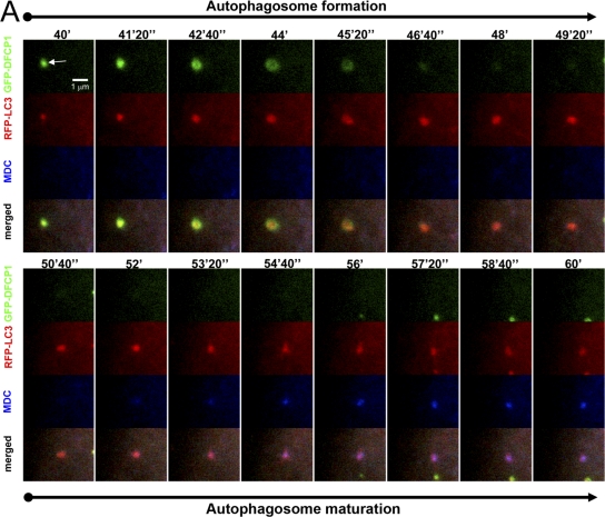
Figure 7.
Autophagosome maturation after omegasome exit. Cells expressing GFP-DFCP1 and mRFP-MAP-LC3 were starved and imaged for the indicated time interval. At 30 min after starvation, the cells were also incubated with 2 μM MDC. Note that an autophagosome emerges first from an omegasome (panels labeled autophagosome formation; arrow indicates the omegasome) and then begins to stain with MDC (panels labeled autophagosome maturation) without appearing to change its appearance or to fuse with another MDC-positive vesicle.

