Figure 1.
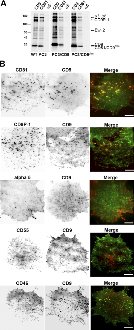
Analysis of tetraspanin assemblies in PC3 cells. (A) Immunoprecipitation experiments in WT PC3 cells or in cells overexpressing CD9 (PC3/CD9) or a nonpalmitoylated form of CD9 (PC3/CD9plm). Biotin-labeled cells were lysed in Brij97 and incubated with anti-CD9, anti-CD81, or anti-α5 antibodies (the latter is used as a negative control). Immunoprecipitated proteins were detected using peroxidase-coupled streptavidin. (B) Immunofluorescence images of PC3/CD9 living cell basal membrane by TIRF microscopy at 37°C. Cells were incubated with the anti-CD9 Cy3B-conjugated antibody SYB-1 (middle; green in the merge image) and with various antibodies labeled with Atto647N (left; red in the merge images) and raised against (top to bottom) CD81, CD9P-1, the α5 chain of integrin, CD55, or CD46. Bars, 10 μm.

