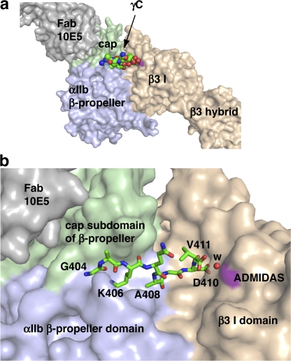
Figure 2.
The binding pocket for the γC peptide. (a) Overview. (b) Detail. αIIbβ3 and 10E5 Fab are shown as solvent-accessible surfaces, with the γC peptide shown in spheres (a) or sticks (b). Lys-406 and Asp-410 are the most buried residues. The surface at the ADMIDAS metal is colored purple (as is that at the MIDAS, which is not visible), and the water coordinating the Val-411 α-carboxyl to the ADMIDAS is a red sphere. Figures were made with Pymol (DeLano Scientific).

