Figure 1.
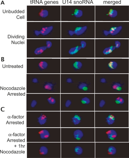
In situ hybridization of unsynchronized and nocodazole-treated cells. In each panel, fluorescent oligonucleotide probes complementary to the U14 snoRNA (green) or 10 tRNALeu(CAA) genes (red) were used for hybridization. Blue represents DAPI staining of nucleoplasmic DNA. (A) Nuclei from unsynchronized cells show that the tRNA gene signal (red) consistently overlaps the nucleolar signal (green) prior to and throughout division of the nucleus. (B) Depolymerization of microtubules by arrest in nocodazole causes partially divided nuclei in which tRNA genes remain clustered, but clusters are divorced from the nucleolus. (C) This effect is not due to nocodozole blockage prior to nuclear division, as demonstrated by the release of tRNA gene clusters from the nucleolus by depolymerization of microtubules in cells arrested in G1 by α factor.

