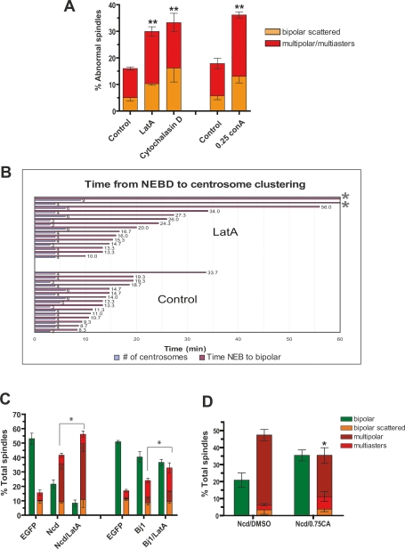
Figure 3.
Actin-dependent forces cooperate with spindle intrinsic forces to cluster supernumerary centrosomes. (A) Actin requirement for centrosome clustering in S2 cells. Cells were treated with Latrunculin (40 μM LatA), Cytochalasin D (20 μM), or Con-A (0.25 mg/mL) for 2 h and the percentage of centrosome clustering defects was determined. Graph shows the average of three independent experiments (mean ± SD, [**] P < 0.005, Student’s t-test). (B) Live cell imaging was used to measure the time from NEBD to centrosome clustering in S2 cells expressing GFP-SAS-6 and mCherry-α-tubulin in the presence or absence of LatA. There is a delay in centrosome clustering in LatA-treated cells (P < 0.02, Student’s t-test), and asterisks indicate the cells that failed to cluster centrosomes (Supplemental Movie S5). (C) Percentage of cells with centrosome clustering defects after RNAi of Ncd or Bj1 (RCC1) alone or in conjunction with LatA treatment (2 h). (D) Calyculin A (0.75 nM for 2 h) partially rescues the centrosome clustering defect in Ncd RNAi-treated cells. Graph shows the average of three independent experiments; mean ± SD. (*) P < 0.05, Student’s t-test. Bar, 10 μm.

