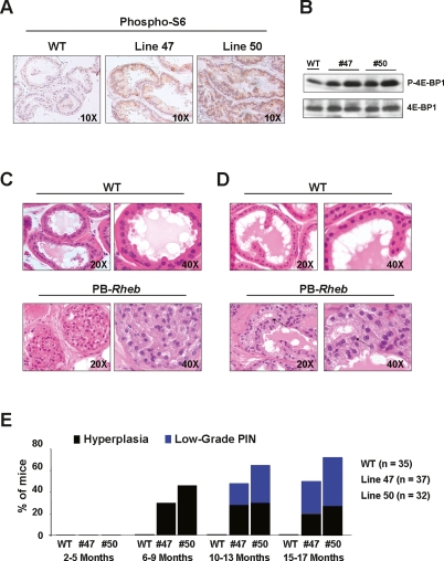
Figure 2.
Analysis of the PB-Rheb mice prostates. (A) IHC staining of prostate sections from wild-type and PB-Rheb mice with an anti-phospho S6 antibody. (B) Western blot analysis of protein lysates from wild-type and PB-Rheb mice. (C) H&E staining of DLP sections from 6-mo-old wild-type and PB-Rheb mice showing hyperplasia in the prostate of the transgenic mice. (D) H&E staining of DLP sections from 10-mo-old wild-type and PB-Rheb mice showing low-grade PIN in the prostate of transgenic mice. An example of nuclear atypia is indicated by arrows. (E) Cumulative incidence of hyperplasia and low-grade PIN over time in wild-type and PB-Rheb mice.

