Figure 6.
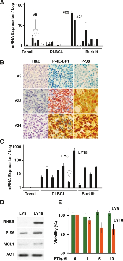
RHEB expression and drug sensitivity in human lymphoma. (A) Quantitative real-time RT–PCR analysis of RHEB expression from cDNAs prepared from reactive tonsils (tonsil), a collection of DLBCL, and some cases of Burkitt’s (Burkitt) lymphoma. (B) Representative micrographs of lymphomas #5, #23, and #24 representing low and high RHEB mRNA-expressing lymphoma in A. Samples are stained with hematoxylin and eosin (H&E), and antibodies against the indicated antigens. (C) Quantitative real-time RT–PCR analysis of RHEB expression in human lymphoma cells lines representing DLBCL and Burkitt’s lymphoma compared with reactive tonsils. (D) Immunoblot of lysates prepared from low (LY8) and high (LY18) RHEB-expressing DLBCL lines probed with the indicated antibodies. (E) Mean and standard deviation of viability of LY8 and LY18 human lymphoma lines treated with FTI-277 at the indicated concentrations for 48 h.

