Figure 2.
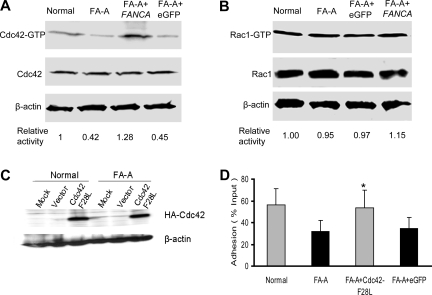
Decreased Cdc42 activity in FA-A cells. (A) Levels of the active, GTP-bound Cdc42 (top panels) in lymphoblasts were determined by the effector-domain (GST-PAK1) pull-down assay followed by Western blotting using an antibody against Cdc42. The relative levels of active Cdc42 are indicated below each pull-down blot. The levels of total Cdc42 (middle panels) and β-actin (bottom panels) are shown as loading controls. (B) Levels of the active, GTP-bound Rac1 in lymphoblasts as determined by the effector-domain pull-down assay followed by Western blotting using an antibody against Rac1. The levels of total Rac1 and β-actin are shown as loading controls. (C) Expression of the constitutively active mutant of Cdc42 (Cdc42-F28L) containing an N-terminal HA tag in normal and FA-A lymphoblasts was analyzed by anti-HA Western blotting. (D) Forced expression of an active Cdc42 enhances adhesion of FA-A lymphoblasts. Untransduced, eGFP (vector)–transduced, or Cdc42-F28L–transduced normal or FA-A lymphoblast cells were subjected to adhesion assays. Data represent the mean plus or minus SD of 3 independent experiments in duplicates. *Difference between the Cdc42-F28L–transduced and untransduced (FA-A) or eGFP-transduced FA-A lymphoblasts is significant at P < .05.

